If you need to print this PDF or save for later
click below to download your copy.
[button size=”medium” style=”secondary” text=”Download PDF” link=”https://vetovation.com/wp-content/uploads/2021/07/Pain-Management-After-Orthopedic-Procedures.pdf” target=”none”]
By Cathy T. Mann
RVT, VTS (Surgery) Surgery and Anesthesia Nurse, Veterinary Specialty Hospital of the Carolinas, Cary, NC
General practice veterinarians often refer clients and patients to specialty surgery centers or veterinary teaching hospitals for various orthopedic procedures including but not limited to tibial plateau leveling osteotomy, fracture repair, and arthroscopy. However, with the advent of traveling board certified veterinary surgeons, many of these procedures are being performed with veterinary orthopedic surgeon equipment in the general practitioner’s clinic, and he/she is now managing postoperative care for patients who underwent procedures performed by someone else.
Successful recovery from orthopedic procedures is typically much more complicated than recovery from routine soft tissue surgeries. Important aspects of recovery from orthopedic procedures may include administration of multiple oral medications, activity restriction, diet adjustment, bandage care, and physical rehabilitation, in addition to the more familiar suture removal, incision care, and Elizabethan collar (e-collar) use. Multiple rechecks and adjustments are common. Clients often call back for advice pertaining to routine care and unexpected complications. This article is intended to provide insight to assist veterinary professionals in these situations.
Discharge Appointments
The key to successfully managing postoperative recovery is communication with the client. Providing detailed, written discharge instructions is essential. A comprehensive discharge appointment should schedule adequate time for the clients to read the discharge instructions and get answers to any questions they may have. A designated nurse should show the client radiographs of the procedure, explain medications and dosing schedules, review home care instructions, demonstrate physical rehabilitation techniques, and help to carefully load the patient into the client’s vehicle for the ride home.
Once clients have read the discharge instructions, they should be shown the postoperative radiographs. Doing so gives the client a visual reference and facilitates a discussion of both the procedure that was performed and the bone healing process. Bone healing takes an average of 2 to 12 weeks, depending on the injury, procedure, and type of repair.1 In some cases, such as a complicated fracture in a geriatric patient, healing can take up to 5 months to a year. Patient factors such as age, nutrition, metabolism, and general health status also affect the healing process.1 Young, healthy puppies generally heal faster than older dogs or dogs with systemic disease. Initial soft tissue wound healing is much faster.2 The skin incision will heal first, and clients should be instructed to return for suture removal in 10 to 14 days. However, the patient’s hair coat takes the longest to return to normal. The outline of the shaved area is often still visible to varying degrees when the patient returns for its 4- and 8-week recheck appointments (FIGURE 1). The hair shaved for an epidural can take longer to grow back completely and may also grow back a different color. Patients with thyroid or other endocrine disease take even longer to heal and to regrow their hair coat.2 Full show coats may take up to a year or more to be restored following a surgical shave.

Recheck appointments are typically scheduled at 2, 4, and 8 weeks (TABLE 1). Skin sutures or staples are removed at the 2-week recheck appointment. The patient’s gait, comfort level, and progress are evaluated at the 4-week recheck. Radiographs are taken at 8 weeks to assess bone healing and implant performance, condition, and location. Radiographs may be taken earlier for young animals or if problems arise.
| Days Since Surgery | Activities and Rechecks |
|---|---|
| 1 to 3 | Massage + PROM + cryotherapy, crate rest |
| 3 to 5 | Incision cover removal, fentanyl patch removal, massage + PROM + heat therapy |
| 10 to 14 | Suture or staple removal +/- aquatic therapy |
| 14 to 28 | Create rest +/- very short leash walks (5 to 10 min) |
| 28 | 4-week recheck with surgeon |
| 28 to 60 | Increased duration of leash walks |
| 60 | 8-week recheck with surgeon, radiographs |
| Over 60 | Gradually increased exercise |
PROM = passive range of motion
Medications
After the procedure has been discussed, then the prescribed medications can be reviewed. Clients should be educated on how each medication is intended to be used and the benefit each provides for the patient. They should be instructed to watch for any adverse effects or interactions between these and any other medications that their pet is taking. Dosing schedules should be relayed, including when the next dose of each medication is due. Clients should be advised on the best technique for administering pills or liquids. The discharging nurse should reinforce the need to refrigerate or shake some products prior to each use and take the time to demonstrate unusual instructions such as how to measure liquid meloxicam or administer buprenorphine transmucosally. Routine heartworm and flea preventatives do not need to be discontinued or delayed as long as no complications have been encountered; however, topical flea and tick preventatives should not be applied to shaved areas or near incision sites. Clients should be advised to stop all prescribed medications and call their veterinarian immediately if any adverse reactions are observed or if they encounter any problems associated with medications.
Although antibiotics are not warranted after clean orthopedic procedures, they are occasionally prescribed for patients with open fractures or concurrent skin infections.3 Elective procedures may be delayed to allow for skin infections to be treated and cleared, but other procedures such as fracture repair cannot always be delayed. Staphylococcus species, Streptococcus species, Escherichia coli, and Pasteurella species are the bacteria most commonly cultured from bone and joint infections. Cephalexin or clindamycin is frequently prescribed following orthopedic surgery since each is very effective against these types of infection.4 However, if an active infection is suspected, the site should be sampled and the sample submitted for culture and sensitivity testing to identify the organism involved and the most effective antibiotic. The surgeon may prescribe an interim antibiotic to be given until these test results are obtained. Common adverse effects of antibiotic use are nausea, inappetence, vomiting, or diarrhea.5 If any of these signs are observed, the clients should call their veterinarian immediately. In some cases, the antibiotic may be replaced. In severe cases, the patient may need to be seen by a veterinarian and treated accordingly.
Typical medications prescribed following orthopedic procedures include nonsteroidal anti-inflammatory drugs (NSAIDs) such as carprofen, meloxicam, robenacoxib, deracoxib, firocoxib, or grapiprant. NSAIDs are used to reduce pain and inflammation. Adverse effects include stomach upset, nausea, vomiting, diarrhea, and gastrointestinal ulceration. NSAIDs should never be given in conjunction with steroids such as prednisone, prednisolone, or dexamethasone because the combination greatly increases the risk for gastrointestinal ulceration.5 It is typically recommended to give NSAIDs and antibiotics with food to minimize any stomach upset associated with these drugs.
In addition to NSAIDS, pain relievers such as buprenorphine, gabapentin, or tramadol may be prescribed. Buprenorphine is a liquid injectable opioid medication that can also be given via the oral transmucosal route. It is more commonly prescribed for cats but is also used for dogs. The higher salivary pH of cats allows for higher levels of absorption and greater effectiveness of the drug.6 When administering this medication, the objective is not for the patient to swallow the dose; instead, the liquid should be applied to the oral mucous membranes of the gingiva, buccal mucosa, or sublingual region for maximum effectiveness. Gabapentin is an analogue of the inhibitory neurotransmitter GABA and is useful as both an anticonvulsant and an analgesic. As an analgesic, it is used to treat neuropathic pain in dogs and cats. Adverse effects include sedation and ataxia, but gabapentin is safe to give with NSAIDs, opioids, phenobarbital, and bromide.5 Tramadol is a weak opioid but has no beneficial effects on signs of pain and orthopedic dysfunction in dogs with osteoarthritis, probably because dogs produce very little of the active metabolite O-desmethytramadol during their metabolism of tramadol.7 At high doses, tramadol may cause seizures in dogs.5 Clients may observe some sedation associated with these and other drugs. If a patient being given any of these drugs becomes overly sedated, this can be addressed by reducing to the minimum effective dose.
An anxiolytic such as trazodone or a sedative like acepromazine is commonly given for especially anxious or overly active patients. Some sedation is acceptable, but the patient should be alert enough to be able to eat regular meals, to move about in its crate or bed, and walk outside to relieve itself. Soiled bedding should be changed immediately to avoid urine scald or fecal contamination of the surgical incision site. Wet or soiled bandages should be changed as soon as possible. When walking, the patient should be able to do so with limited assistance and without stumbling or falling due to excessive sedation. Stumbling or falling may result in further injury or failure of the recent repair. If a patient is unsteady, it can be assisted by a harness for front limbs or a towel sling for rear limbs. Many commercial products are available for this type of assistance. The Web Master harness assists with front limbs, the Quick Lift or GingerLead assists with hind limbs, and the Help ’Em Up harness assists with both front and hind limbs. Harnesses should be adjusted to be snug enough to provide support without being uncomfortable. Steady the pet by holding the harness near the shoulders or by the handle provided rather than using an attached lead. Slings should be placed just in front of the hind limbs at the patient’s waist. Hold the sling to provide support by steadying the pet without carrying the hind limbs.
Fentanyl patches are often used following orthopedic procedures and deliver a constant dose of pain medication via the transdermal route. Common locations for fentanyl patches include the metatarsals, metacarpals, the nape of the neck, or over the epaxial muscles. As with the epidural site, regrowth of the hair coat at the site of fentanyl patch placement may also be sporadic and discolored. Before placing a patch, prepare the site by shaving the hair, being careful not to abrade the skin. Remove any loose hair by wiping with dry gauze or using a lint roller. If the site is dirty, it can be gently wiped clean with saline or warm water and allowed to dry prior to patch placement. Alcohol or a detergent cleanser should not be used as these products disturb the lipid matrix of the stratum corneum, resulting in delayed absorption of the drug.8 The patch is applied directly to the skin and covered with a tape bandage labeled with the fentanyl concentration, date and time applied, date to remove, and initials of the person who applied the patch. The drug contained in the patch may take up to 12 to 24 hours to reach therapeutic plasma concentration and is then delivered at a constant rate over the course of 3 to 5 days.8 A specific removal date should be stated in the discharge instructions. Adverse reactions can include bradycardia, sedation, anorexia, dysphoria, and skin reaction at the placement site.8 The FDA recommends disposing of patches by folding them in half with the sticky sides together and then flushing them down the toilet. They should not be placed in the household trash where children or pets can find them.9 However, flushing of patches is discouraged or prohibited in some states due to environmental concerns. The client may choose to return to the clinic for patch removal and disposal.
Incision Care
Removal of the fentanyl patch can coincide with the removal of a simple wound cover applied the day of surgery. The simple wound cover typically consists of a primary layer of triple antibiotic ointment, a secondary layer of a nonadherent dressing, and an adhesive covering such as Hypafix (FIGURE 2). The wound cover helps keep the surgical wound clean and free of contamination. Once it is removed, the client should monitor the incision for signs of infection or damage. Instruct clients to look for redness, swelling, discharge, and missing sutures or staples. A small amount of clear or serosanguinous discharge within the first day of surgery is normal but should stop by the time the wound cover is removed. Any discharge that is thick, bloody, purulent, white, yellow, green, or foul smelling may indicate a problem and should be rechecked by the surgeon. Missing sutures or staples indicates self-mutilation or trauma to the wound and should also be evaluated by the surgeon. An e-collar should be utilized to prevent further damage. Client and patient compliance may need to be investigated if damage has occurred with the collar in place.

An e-collar should be provided to all postoperative patients to provide a physical barrier against the wound or bandage. Mutilation of a bandage can result in the cost of bandage replacement, ingestion of bandage material and possible intestinal obstruction, and access to the surgery site. Licking and chewing of the surgery site can result in suture or staple removal, disruption of the wound and repair, delayed healing, and surgery site infections. Various products on the market are available to clients who wish to use a different product to achieve the same results. Softer versions such as the Comfy Cone, Recovery Collar, and ZenCone are available through pet stores and online retailers and can be gentler to the pet, the client, and the household. Alternatives such as the inflated Cloud Collar and the BiteNot collar aim to prevent patients from bending their neck enough to reach the surgery site and can be used effectively for certain pets. An inexpensive version of the BiteNot collar can be improvised by using a folded towel taped around the patient’s neck.10 This may be useful for patients that are prone to moist dermatitis caused by drool, water, or wet food, which can become trapped inside nonporous e-collars. The towel can also be replaced regularly and washed. Instruct clients that some pets may be quite averse to certain activities such as urinating, defecating, eating, and drinking while wearing an e-collar or alternative. It can certainly be removed during these activities and whenever the pet is being directly supervised but must be worn whenever the pet cannot be observed, including during the day when the clients are at work and overnight while they are asleep.
Bandage Care
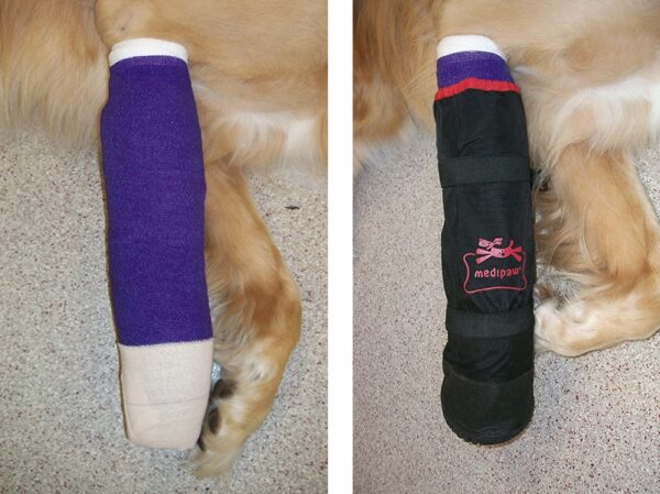
External coaptation devices provide additional challenges and responsibilities for the patient care provider. Splints and bandages are most commonly associated with fractures or wounds that occur distal to the elbow or stifle joint. Clients should be instructed to monitor the condition of such appliances or bandages. If the patient’s toes are accessible, they should be checked for signs of heat, cold, or swelling. The client should be taught how to inspect and feel the toes before leaving the clinic so that they have a baseline for comparison. The bandage should be protected with a waterproof covering when the pet goes outdoors. A plastic bag, a modified IV fluid bag, or a Medipaw can be used for this purpose (FIGURE 3). However, to prevent condensation, the covering must be removed when the pet is indoors. The bandage should be checked by the surgeon or attending veterinarian if any of the following conditions occur: the bandage slips or gets wet, the patient suddenly stops bearing weight on that limb, the patient damages the bandage, any foul-smelling odor is coming from the bandage, or the bandage falls off.10 Clients should never attempt to modify or replace a bandage at home. Barring complications, bandages should be changed and the limb evaluated on a regular basis, as often as every 3 to 5 days but no longer than every 10 to 14 days. This timing varies greatly, depending on the nature of the repair and the condition of both the bandage and the patient’s skin beneath it. It is recommended that the first bandage change appointment be scheduled at the time of discharge and that each subsequent appointment be scheduled during the current appointment.
Home Care
Occasionally, clients become concerned that their pet is off its usual schedule of eating, drinking, urinating, or defecating. Some variation can be expected but can also escalate to be problematic. Some pets refuse to eat in the hospital but will regain their appetite once returned to their home environment. Others may remain picky eaters at home. There are a few tricks that can be used to induce them to eat. Low-sodium or no-sodium varieties of gravy, bone broth, or cottage cheese can be added to make food more palatable. Dogs may also like honey or maple syrup, but sweet flavors are not preferred by cats. Putting pet food on a regular dinner plate first placed on the table and then offered to the pet may trick the pet into thinking it is getting special treatment.11 Low-sodium bone broth can also be added to the water bowl to encourage increased water intake. Clients can temporarily cook specifically for their pet. If this is the case, they should consult their veterinarian for recommendations to ensure they are preparing a complete and balanced diet.
If an epidural was administered prior to surgery, the patient may need to have its bladder expressed or catheterized if it has not urinated within 24 hours of surgery, but it should regain bladder control before being discharged.12 A patient may not urinate or defecate for several reasons. Dogs that are normally off leash may be shy when leashed walked. They may also be substrate-specific, meaning they like to go on grass, near bushes, or in the woods. Try walking them calmly to their preferred areas. If the favorite area is far away, a small dog can be carried to the area and then allowed to choose his spot. Covered litter boxes should be uncovered or replaced with low-sided trays that are easier to access. Cats may not appreciate their clay litter being replaced with shredded paper. Try using Yesterday’s News or other alternative litter. If this is unacceptable to the cat, then compromise with a very shallow layer of nonclumping clay litter. In some cases, a male dog will be very reluctant to bear weight on the surgery leg if he is accustomed to lifting the contralateral limb to urinate. Female dogs may be reluctant to squat if surgery involved the hind limb or pelvis. Ensure that their pain is well controlled and that they are able to posture to urinate or defecate. Injuries involving the pelvis may result in narrowing of the pelvic canal. In some of these cases, a stool softener may be required. Some delay before defecation is quite common. Patients are fasted before surgery and are administered narcotics perioperatively. Narcotic use can result in both inappetence and reduction of gastrointestinal motility.5 As a result, it may take a few days for the body to produce a bowel movement. If the pet is straining to urinate or defecate or having diarrhea, then it may need to be seen by a veterinarian.
Activity Restriction
In the first few weeks, the patient should be restricted to a crate or small room when not being directly supervised but can be allowed out when directly supervised. Cats should be discouraged from jumping up to and down from high levels. They can be kept in a large dog crate while unsupervised. Avoid slippery surfaces such as hardwood, tile, or vinyl flooring. Inexpensive indoor/outdoor carpeting can be purchased from home improvement stores and used to temporarily cover these surfaces. Bathroom rugs that have nonskid backing can be used in crates, bathrooms, or laundry rooms. Baby gates can be utilized to block doorways to restricted areas. If the pet is being kept in a bathroom or laundry room, use of a baby gate rather than a closed door allows the pet to be monitored and less isolated from the family. Be sure to pet-proof the room as well. Dogs can be given puzzle toys to occupy them while restricted. Broth or diluted peanut butter can be placed in a fillable Kong toy and frozen before being given to a dog. Avoid activities such as running, jumping, playing with other dogs or people, and navigating full flights of stairs. If stairs are unavoidable, either carry the pet or use a sling and ensure that the pet is using the stairs carefully. Do not allow the pet to charge up and down or jump over the stairs. Keep in mind that weight bearing increases on the front limbs when going down and increases on the hind limbs when going up stairs and inclines.
Although some activity restriction is appropriate, cage rest alone is not recommended. The goal of recovery following an orthopedic repair is for the bone to heal and return to normal function without implant failure. A gradual increase in weight bearing activity combined with physical rehabilitation is used to ensure success. Lack of activity results in muscle atrophy and disuse osteopenia. Muscles must be strong and utilized in order to assist the weight bearing of the bones without overexerting stress on the metal implants.13 The increase of pace and intensity of activity will depend on the patient as well as the stability of the repair. The key to success is gentle, controlled walks at a slow enough pace to encourage weight bearing. Low impact exercises such as walking on flat surfaces are the first stage to recovery. Begin with short leash walks several times daily to allow the pet to urinate and defecate. Clients often ask if they will need to motivate their pet to be more active after orthopedic surgery. The opposite is more often true. Pets will typically want to be more active much sooner than desired. If the patient has an abundance of energy that needs to be burned off, then more frequent short walks are preferable to longer walks. Once sutures are removed, walk lengths can be extended gradually, being careful to avoid overexertion. It is recommended to allow at least a 2-hour break between exercise sessions. If the pet seems tired or sore after activity, then the next session should be shorter. Again, frequency can be increased to compensate for the shorter duration of activity. As the pet improves, duration can be extended and intensity can be increased by walking on inclined or uneven surfaces. Controlled use of stairs can also be added. These levels of activity should be discussed during the 2-, 4-, and 8-week recheck appointments (TABLE 1). If at any time the patient displays a sudden change in use of the surgery limb, such as increased limping or no longer weight bearing, then the patient should be checked by the surgeon as soon as possible.
Physical Rehabilitation
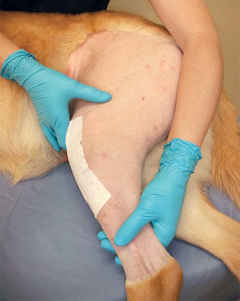
Passive range of motion (PROM) and massage are excellent techniques that can be started shortly after surgery, taught to the client during the discharge appointment, and utilized at home. Begin with the pet lying down with the affected limb up. If the pet objects to lying down, it can stand. Gently rub the muscles associated with the affected limb. This relaxes the pet and loosens the muscles that will be worked during PROM. A second person can help by petting the animal’s head, rubbing its ears, and providing verbal reassurance. Treats can be given for positive reinforcement, but the regular diet will need to be adjusted accordingly. Alternatively, pieces of dry kibble can be used. Continue massage until the pet is relaxed. Make sure to support the limb appropriately, placing your hands above and below the joints, not on the joints. The forelimb can be held just below the shoulder and above the carpus. The hind limb can be held above the knee and just above or below the tarsus (FIGURE 4). Hold the limb parallel to the floor in its natural weight-bearing position. Be careful not to abduct or adduct the limb into an unnatural position or place any torque on the lower part of the limb. Guide the entire limb through flexion and extension of each joint to mimic the motion of walking. If the patient resists completely, then return to massage for a bit longer and try again. Full extension and flexion is unlikely to be achieved in the first PROM session, but over time and multiple sessions, improvement should be achieved. Repeat up to 10 to 15 cycles for each session. The goal is to do 2 sessions per day, but sessions can be increased to as often as every 4 hours.
Cryotherapy should be done at the end of each session. Clients can create their own icing bags by mixing 1 part isopropyl alcohol with 2 parts water in a plastic freezer bag. Double-bag the contents and freeze for at least 1 hour between sessions. The mixture will be cold but will remain pliable to conform to the animal’s body shape. Wrap the ice pack in a towel before applying it to avoid damaging the skin. Keep the ice pack in place for 15 to 25 minutes at the end of each session. Two to three days after surgery, switch from ice packs to warm packs. Warm packs can be made by placing dry rice or beans in a bag or a sock and warming them in the microwave. Clients must be extremely careful when heating these packs because if too hot, they can cause serious burns. Instruct clients to wrap the warm pack in a towel before use and to check the pack on themselves before applying it to their pet. Once the pet is reliably bearing weight while walking, PROM can be discontinued.
After the incision has healed, typically after suture or staple removal, then swimming is an approved exercise. The patient’s comfort level with water and level of fitness must, of course, be taken into consideration before starting aquatic therapy. Despite the advantages of buoyancy, swimming is strenuous aerobic exercise and clients should be warned not to let their pet overexert itself. Swimming allows for a greater range of motion of the shoulders and elbows and provides excellent muscle exercise while minimizing pain by reduced weight bearing.13 However, swimming is more beneficial for the front limbs than for the hind limbs. Patients recovering from hind limb procedures may benefit more from hydrotherapy in an underwater treadmill under the supervision of a veterinary rehabilitation professional. The underwater treadmill has several advantages, such as the ability to vary water levels, jet resistance, and tread speed.13 The clinician can also provide assistive techniques to increase range of motion or ensure proper joint movement and can reassure a patient that is not comfortable with the water.13
Nutrition
All patients should be fed a complete and balanced diet. No specific additions are required, but some clients may elect to add certain supplements, such as glucosamine and chondroitin sulfate. These supplements are intended not to aid healing but rather to prevent or slow the effects of osteoarthritis. Because recovery often results in a significant decrease in activity level, some patients may benefit from dietary restriction as well. Typically, a 10% to 20% decrease in calories is adequate to avoid weight gain during recovery. This is particularly important for overweight and obese patients. Increased weight exerts more stress on implants and is an additional hurdle to recovery.13 Once activity level is restored, the diet can return to normal. Obese patients may require additional visits to the veterinarian to determine the appropriate diet plan for recovery and beyond. If weight loss cannot be achieved with reduced calories, a prescription diet may be warranted.
Summary
Important aspects of orthopedic recovery include administration of multiple oral medications, especially analgesics; activity restriction; bandage care; physical rehabilitation; and diet adjustment. All of these can be successfully managed by good communication with the client, beginning at the time of discharge and continuing with multiple recheck appointments throughout the recovery process.
References
- DeCamp CE, Johnston SA, Dejardin LM, Schaefer SL. Brinker, Piermattei, and Flo’s Handbook of Small Animal Orthopedics and Fracture Repair. Saint Louis: Elsevier; 2016:35-37.
- Swaim SF, Krahwinkel D. Management of hard to heal wounds. Veterinary Clinics of North America: Small Animal Practice, Wound Management. Philadelphia: W.B. Saunders; 2006:707-708.
- Bergh MS. Antimicrobial regimens and surgical site infections. Clinician’s Brief 2016;14(3):17-20.
- Windahl U, Bengtsson B, Nyman AK, Holst BS. The distribution of pathogens and their antimicrobial susceptibility patterns among canine surgical wound infections in Sweden in relation to different risk factors. Acta Vet Scand 2015;57:11.
- Papich MG. Saunders Handbook of Veterinary Drugs: Small and Large Animal. 4th ed. Saint Louis: Elsevier; 2016:143, 218, 321, 349-350, 543, 802, 805.
- Robertson SA, Taylor PM, Sear JW. Systemic uptake of buprenorphine by cats after oral mucosal administration. Vet Rec 2003;152(22):675-8.
- Hofmeister EH, Egger SM. Transdermal fentanyl patches in small animals. JAAHA 2004;40:468-478.
- Budsberg SC, Torres BT, Kleine SA, Sandberg GS, Berjeski AK. Lack of effectiveness of tramadol hydrochloride for the treatment of pain and joint dysfunction in dogs with chronic osteoarthritis. JAVMA 2018;252(4):427-432.
- U.S. Food and Drug Administration. Fentanyl patch can be deadly to children. https://www.fda.gov/ForConsumers/ConsumerUpdates/ucm300803.htm#2 (accessed May 15, 2018).
- Swaim SF, Renberg WC, Shike KM. Small Animal Bandaging, Casting, and Splinting Techniques. Ames: Wiley-Blackwell; 2011:10, 113-114.
- Linder D. How do I get my picky pet to eat? http://vetnutrition.tufts.edu/2017/03/how-do-i-get-my-picky-pet-to-eat/ (accessed May 15, 2018).
- Steagall PVM, Simon BT, Teixeira Neto FJ, Luna SPL. An update on drugs used for lumbosacral epidural anesthesia and analgesia in dogs. Frontiers in Veterinary Science 2017;4:68.
- Zink MC, Van Dyke JB. Canine Sports Medicine and Rehabilitation. Ames: Wiley-Blackwell; 2013:74, 75, 159.